Urgent Help Needed: Undiagnosed Condition in 8-Month-Old with Severe Symptoms
Hello everyone! I'm posting on behalf of a friend in China whose 8-month-old son has been battling severe, unexplained symptoms since he was just 2 months old. These symptoms include [translated] congenital aneurysm, lymphangioma, bone defect, old thrombus in the abdominal aorta, carotid aneurysm, abnormal shape of both kidneys and inferior mesenteric artery, invisible and patchy bone at the distal end of the right condylar artery, and sepsis outbreak. The family has consulted top pediatricians and hospitals across Beijing, Jinan, Shanghai, and more but has been unable to get a definitive diagnosis. Genetic testing has revealed no clear genetic issues.
If anyone has information, resources, or insights on these symptoms or their potential causes, your help would be deeply appreciated by the family. Thank you!
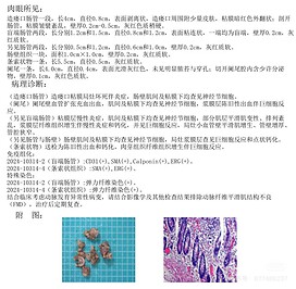
Here is the translated pathology report the family received last week (week of October 21):
What we see with our naked eyes: A section of intestinal tube at the stoma was 4cm long and 0.8cm in diameter, with a peeled surface, a small amount of skin attached around the stoma, and dark red mucosa everted; when the intestinal tube was opened, the mucosal folds were disordered, the wall thickness was 0.2cm-0.5cm, and the gray-red texture was slightly hard. There are two sections of blind-ended intestine, 1.2 cm and 1.5 cm long, 0.8 cm and 1.2 cm in diameter, with adhesion on the surface, one end is blind, the wall is 0.2 cm thick, and the texture is gray-red and soft. There are also two sections of intestinal tube, 0.5cm and 1.2cm long, 0.6cm and 1.0cm in diameter, 0.2cm thick, gray-red and soft. There is also a piece of intestinal wall tissue, 1.0cm×1.0cm in area, 0.2cm thick, gray-red and soft. A cord-like object, 5.5cm long, 0.5cm in diameter, gray-red and soft. There was one appendix, 4.0 cm long and 0.4 cm in diameter, with a smooth gray-red surface and no obvious pus or perforation. The appendix cavity contained a small amount of secretions, with a wall thickness of 0.1 cm and gray-red and soft texture. Pathological diagnosis: (Stomal intestinal tube) Focal necrosis and inflammation of the stoma mucosa, ganglion cells were found in the intestinal wall muscle and submucosal. (Appendix) The blood vessels in the appendix wall were dilated, congested and hemorrhagic. Ganglion cells were found in the intermuscular and submucosal layers. There was old hemorrhage in the serosa layer with giant cell reaction. (See also blind-end intestinal tube) Chronic inflammation of the mucosal layer, ganglion cells are found in the myenteric and submucosal regions, some smooth muscle degeneration and disordered arrangement, fibrous tissue hyperplasia of the serosa accompanied by chronic inflammation and calcification, giant cell reaction, smooth muscle hyperplasia of focal small blood vessel walls, thickening of the vessel walls, and stenosis of the lumen. (See also Intestinal Tube and Intestinal Wall) Ganglion cells were found in the intestinal wall muscle and submucosal layer, and giant cell reaction and punctate calcification were found in the focal serosa. The cord-like structures were sent for examination and showed old hemorrhage and calcification, granulation tissue, fibrous tissue proliferation and giant cell reaction. Immunohistochemistry: 2024-10314-2 (blind intestinal tube): CD31 (+), SMA (+), Calponin (+), ERG (+). 2024-10314-4 (cord-like tissue): SMA(+), ERG (+). Special staining: 2024-10314-2 (blind intestinal tube): elastic fiber staining (+). 2024-10314-4 (cord-like tissue): elastic fiber staining (+). Consider arterial dysplasia in combination with clinical findings. Please rule out arterial fiber muscle dysplasia (FMD) in combination with imaging and other examination results, and have regular follow-up examinations after treatment. Attached pictures:
